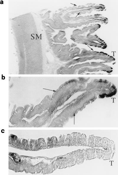
FIG. 1.
The xIFABP gene is transcribed in adult Xenopus gut specifically in the differentiating epithelium. Xenopus intestine was analyzed by whole-mount in situ hybridization. Sections of processed tissue are shown. (a) Villus architecture, with smooth-muscle (SM) layers on the left and the villus proximal-distal axis running left to right toward the tip (T). The dark strain indicates the pattern of xIFABP transcripts following development of the alkaline phosphatase reaction to detect the hybridized antisense xIFABP RNA probe. (b) Higher-magnification view of a villus section. Arrows in both panels indicate the positions along the villus axis that transcripts are first detected in the epithelium. The signal increases in intensity along the proximal-distal axis and is strongest at the distal tip (T). (c) Section from similarly processed tissue that was hybridized with a control sense-strand RNA probe.

