Fig. 1.
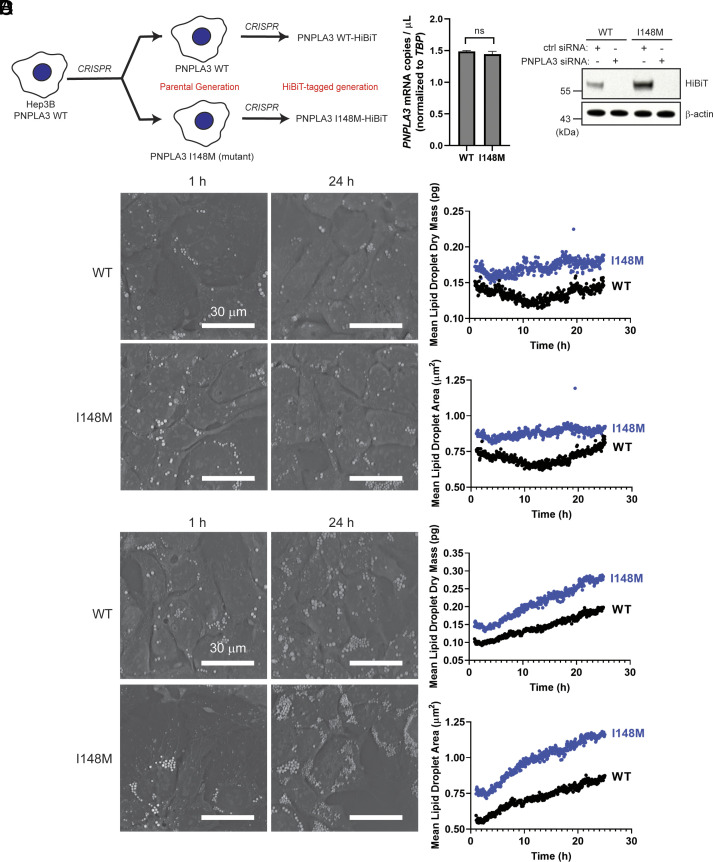
Constitutive endogenous expression of PNPLA3-I148M increases the LD content of hepatoma cells. (A) PNPLA3-I148M was introduced into Hep3B cells using CRISPR. Parental Hep3B cells (WT and I148M) were further modified to introduce a C-terminal HiBiT tag at the endogenous locus. (B) PNPLA3 expression in the parental cells (Left) was analyzed by digital PCR (P = 0.0972, determined by Student’s t test, n = 3). Protein expression (Right) was analyzed by HiBiT detection (bioluminescence observed in the presence of LgBiT protein, as described in SI Appendix) in the tagged cell lines treated with a negative control siRNA or PNPLA3-specific siRNA. (C and D) Representative microscope images (from 1 and 24 h) and quantification of mean lipid droplet content and mean lipid droplet area from live-cell label-free imaging using the Nanolive 3D Cell Explorer microscope. Cells were treated with vehicle (C) or with 200 µM oleic acid (D). Images were taken every 3 min for 24 h, starting 1 h after addition of oleic acid. Quantification was done using Eve software. Experiment was repeated twice with similar results.

