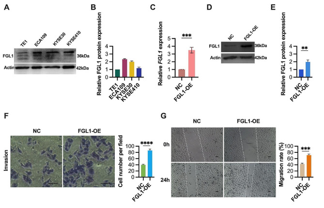
Fig. 2.
FGL1 promotes invasion and migration of ESCC cells. (A-B) FGL1 expression levels were assessed in ESCC cell lines by western blotting. β-Actin served as the loading control. (C-E) FGL1 expression levels were assessed in TE1 cells transfected with FGL1 (FGL1-OE) or random nonsense sequences (NC) using qRT-PCR (C) and western blotting (D and E). (F) The effects of FGL1 overexpression on TE1 cells invasion were evaluated by transwell invasion assay. Scale bars, 100 μm. (G) The effects of FGL1 overexpression on TE1 cells migration were evaluated by wound healing assay. Scale bars, 100 μm. The statistical difference was assessed with two-tailed unpaired Student t test in C, E, F and G. Error bars show the SD from three independent experiments. **p < 0.01, ***p < 0.001, ****p < 0.0001

