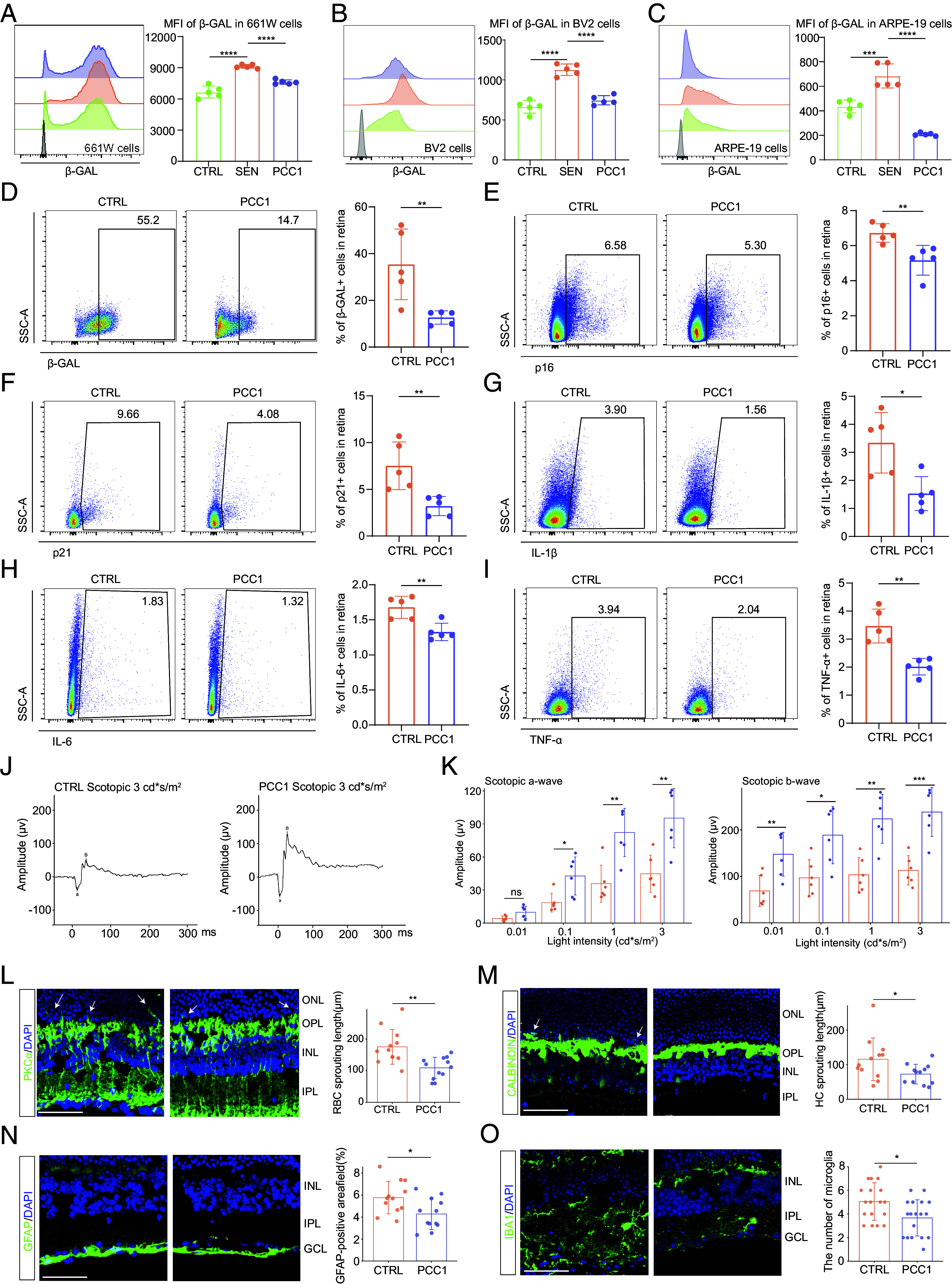
Fig. 2.

Long-term PCC1 treatment relieved functional and structural impairment in the aged retina by both senolytic and senomorphic effects. (A–C) Representative FC histograms (Left) and quantification (Right) of MFI of β-GAL in 661 W (A), BV2 (B), and ARPE-19 (C) cells among three groups (n = 5/group). (D–I) The FC histograms (Left) and column charts (Right) showing the level of β-GAL (D), p16 (E), p21 (F), IL-6 (G), IL-1β (H), and TNF-α (I) in retinal cells between control and PCC1-treated aged mice (n = 5/group). (J) Representative scotopic ERG waveform of aged and PCC1-treated aged mice at a light density of 3 cd*s/m2. (K) Bar charts showing the quantification of scotopic ERG amplitudes (n = 6/group). (L–O) Representative confocal images of retinal frozen sections of aged (Upper) and PCC1-treated (Middle) mice and bar charts of quantification (Right, n = 6/group). Frozen sections are labeled with PKCα (L), Calbindin (M), GFAP (N), and IBA1 (O). Arrows indicate the abnormal sprouting dendrites of RBCs (L) and HCs (M) which extend beyond OPL into the ONL. (Scale bar, 50 μm.) Data are shown as mean ± SD. P values were analyzed using one-way ANOVA with Bonferroni post hoc test (A–C) or two-tailed unpaired Student’s t test (D–I and K–N) or two-tailed Mann–Whitney U test (O); ns, nonsignificant, *P < 0.05, **P < 0.01, ***P < 0.001, and ****P < 0.0001.
