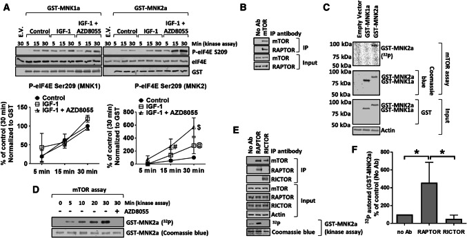
Fig. 1.
MNK2a is a direct substrate for mTOR. a HEK293 cells were transfected with vectors encoding WT GST-MNK2a; 32 h later, cells were starved of serum for 16 h and then transferred to KRB for 30 min. Cells were then treated with IGF-1 for 30 min in the presence or absence of 1 µM AZD8055. GST-MNK1a/2a was then isolated from the lysates on glutathione beads and subjected to MNK kinase assay for the indicated times using recombinant eIF4E as substrate. Assay products were separated and analysed by SDS-PAGE/Western blotting (WB). E.V empty vector. b mTOR was immunoprecipitated (IP) from lysates of HEK293 cells cultured in growth medium. The presence of mTOR and RAPTOR in the immunoprecipitates and input lysates was assessed by SDS-PAGE/WB. c HEK293 cells were transfected with vectors for WT GST-MNK1a or GST-MNK2a; cells were starved of serum for 16 h followed by incubation in KRB for 1 h. Cells were then lysed and GST-MNKs were pulled down and then eluted from glutathione beads. The presence of GST-MNK1a or GST-MNK2a was verified by SDS-PAGE/WB. Eluted GST-MNKs were incubated with immunoprecipitates (containing mTOR) from b for 30 min in assays including [γ-32P]ATP. Radioactivity was detected using a phosphorimager. d mTOR kinase assays were carried out as in c for the indicated periods of time. e mTORC1 or 2 was immunoprecipitated with antibodies to RAPTOR or RICTOR, respectively. Kinase assays were then carried out using [γ-32P]ATP and GST-MNK2a as substrate (for 30 min; with detection by phosphorimager). f Quantification of data in e. Quantification of data in panels a, f is presented as means ± SD. n = 3. *0.01 ≤ P < 0.05 (one-way ANOVA). @0.01 ≤ P < 0.05; #0.001 ≤ P < 0.01; $P < 0.001 (two-way ANOVA)

