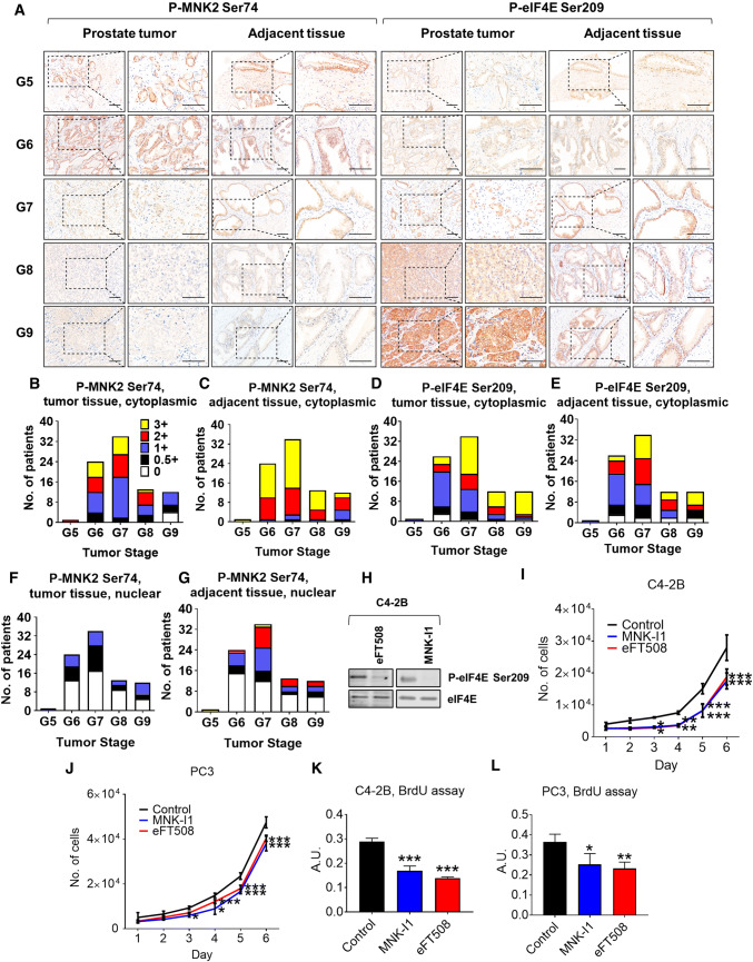
Fig. 7.
Loss of MNK2 Ser74 phosphorylation correlates with advanced prostate tumors in patients. a Levels of P-MNK2 Ser74 in prostate cancer patient samples were assessed by tissue microarray analysis against 84 paired prostate tumors and adjacent normal tissue. Representative IHC images from Gleason score 5–9 (G5–G9) were shown. Scale bar 50 μm. b–i Average immunoreactivity in a was graded in a blinded manner on a scale of 0 (none), 0.5 + (very weak); 1 + (weak); 2 + (intermediate); and 3 + (strong) in b–e: cytoplasmic; f, g: nuclear; b, c, f, g: P-MNK2 Ser74; d, e: P-eIF4E Ser209. b, d, f: tumor tissues; c, e, g: adjacent normal tissues. h C4-2B cells were treated with vehicle (control), eFT508 (2 μM) or MNK-I1 (2 μM) for 24 h before lysis and immunoblotting analysis for P- (Ser209) or total eIF4E. C4-2B (i) or PC3 (j) cells were treated with vehicle (control), eFT508 (2 μM) or MNK-I1 (2 μM), the number of cells were counted for the indicated periods of time. C4-2B (k) or PC3 (l) cells were treated with vehicle (control), eFT508 (2 μM) or MNK-I1 (2 μM) for 48 h, before subjected to BrdU incorporation assays. In panels h–l data are shown as means ± SD, n = 3. *0.01 ≤ P < 0.05; **0.001 ≤ P < 0.01; ***P < 0.001 (one-way ANOVA)

