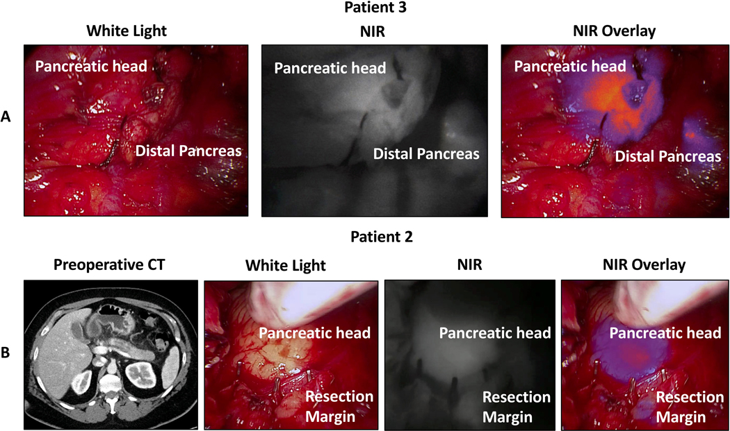
Figure 2:
Intraoperative near-infrared (NIR) imaging from two patients with fluorescence at the initial resection margin. A) In situ intraoperative white light, near infrared, and overlay images after pancreas transection and prior to specimen removal for Patient 3. The patient underwent a pancreaticoduodenectomy and had both fluorescence at the margin and a positive frozen section. The specimen was cut back further to achieve a negative margin. B) Preoperative CT, in situ intraoperative white light, near infrared, and overlay images after pancreas resection for Patient 2. The patient initially underwent a distal pancreatectomy for duct dilation without a clear tumor. Intraoperative imaging showed diffuse fluorescence throughout the pancreas including in the pancreatic head and at the transection margin. The patient had a positive margin on final pathology and eventually underwent a completion pancreatectomy at which time adenocarcinoma was found throughout the pancreatic head.

