
Figure 1.
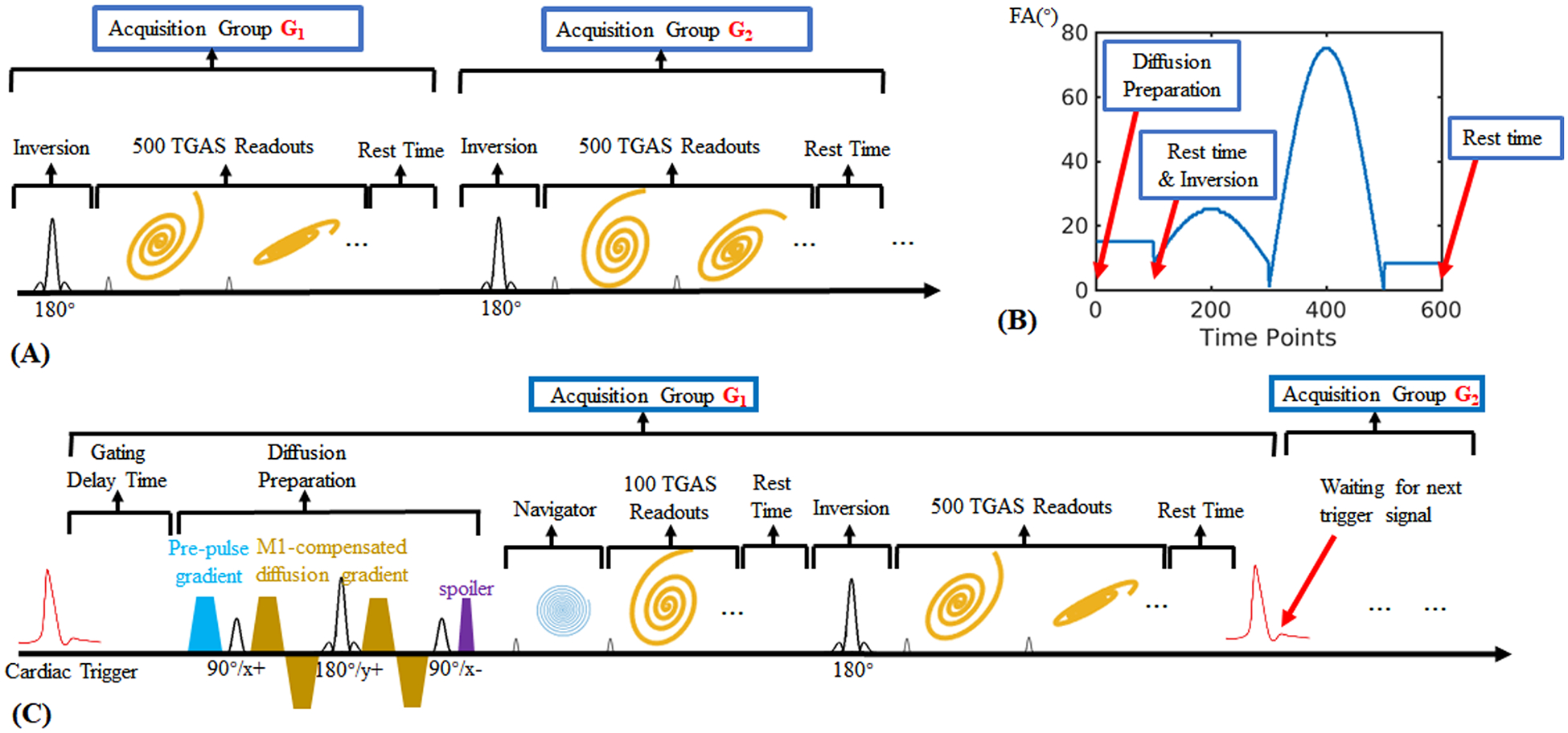
(A) The sequence diagram of a normal 3D MRF based on spiral projection (3D-SPI-MRF) method, which aims to obtain the quantitative T1, T2 and PD maps.
(B) The variation pattern of flip angles used in the proposed DTI-MRF method.
(C) The detailed sequence diagram of the proposed DTI-MRF method.