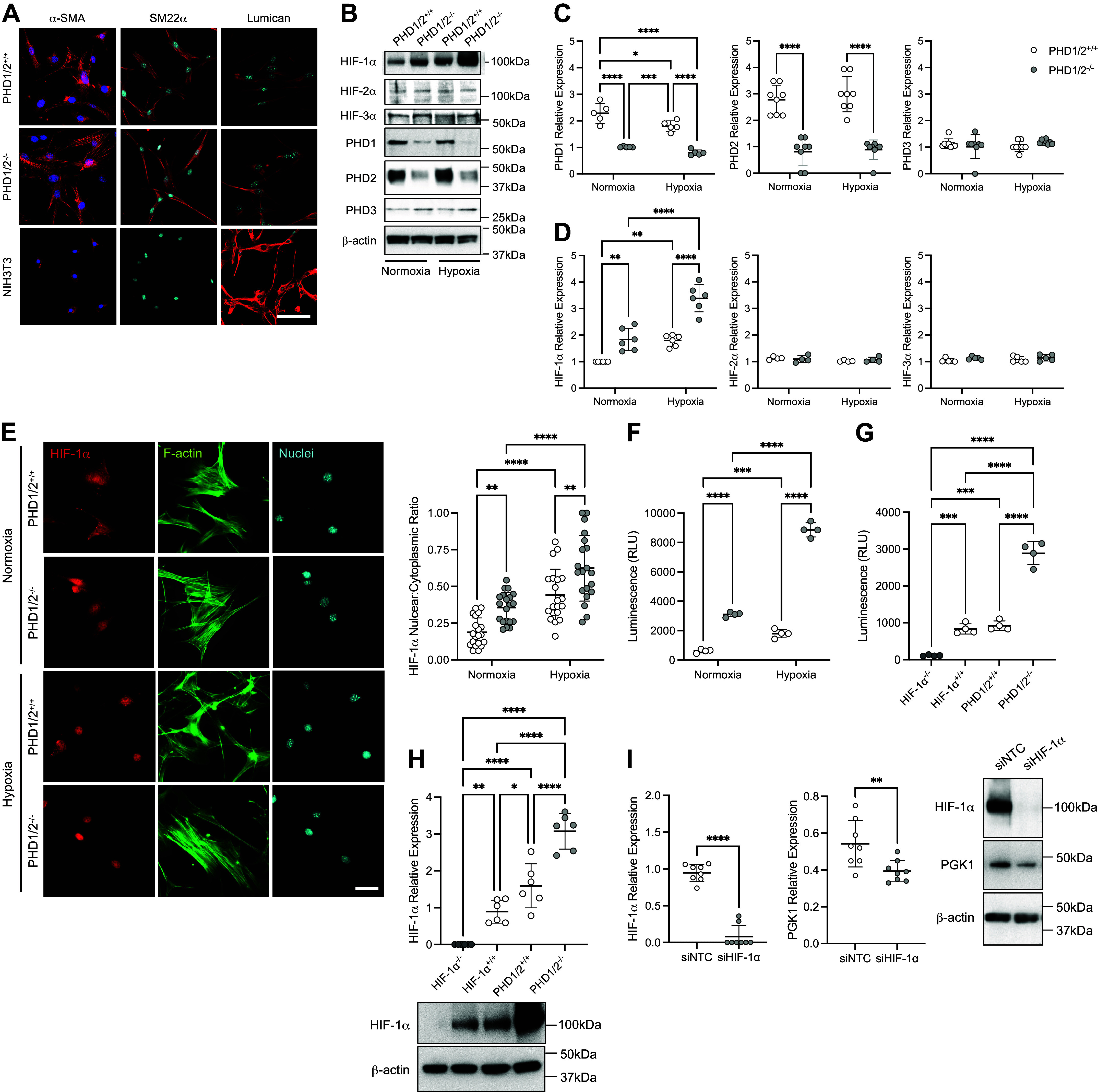
Figure 1.

Characterization of HIF-1α in PASMC isolated from SM22α-PHD1/2+/+ and SM22α-PHD1/2−/− mice. Mice expressing Cre recombinase (Cre+) under the control of the SM22α promoter or without (Cre−) were crossed with PHD1flox/flox; PHD2flox/flox mice to generate SM22α-PHD1/2−/− and SM22α-PHD1/2+/+ mice, respectively. A: expression of α-SMA, SM22α, and Lumican in isolated PASMC (n = 3 replicate experiments). α-SMA (red), SM22α (red), Lumican (red), nuclei (blue); ×100 magnification, calibration bar 100 µm. B: protein expression profile of isolated PASMC exposed to normoxic (21% O2) or hypoxic (1% O2, 1 h) conditions. C: quantification of PHD1, PHD2, and PHD3 expression in isolated PASMC (n = 5–8 replicate experiments). D: quantification of HIF-1α, HIF-2α, and HIF-3α expression in PASMC (n = 4–6 replicate experiments). E: nuclear HIF-1α expression in PHD1/2 PASMC (n = 4 replicate experiments with five fields analyzed for each). HIF-1α (red), F-actin (green), nuclei (blue); ×200 magnification, calibration bar 50 µm. F: HIF-1α transcriptional activity in PASMC under normoxic and hypoxic conditions (n = 4 replicate experiments performed in triplicate). G: HIF-1α transcriptional activity in PASMC isolated from SM22α-HIF-1α−/−, SM22α-HIF-1α+/+, SM22α-PHD1/2−/−, and SM22α-PHD1/2+/+ mice (n = 4 replicate experiments performed in triplicate). H: HIF-1α protein expression in HIF-1α and PHD1/2 PASMC (n = 6 replicate experiments). I: expression of Phosphoglycerate kinase 1 (PGK1), a target of HIF-1α, in siHIF-1α-transfected PHD1/2−/− PASMC (n = 8 replicate experiments). Graphs represent the means ± SD, *P < 0.05, **P < 0.01, ***P < 0.001, ****P < 0.0001, as indicated. P values were measured by two-way ANOVA (C, D, E, F), one-way ANOVA (G, H), and unpaired t test (I). HIF-1α, hypoxia-inducible factor-1α; PHD, prolyl hydroxylase domain; PASMC, pulmonary artery smooth muscle cell; SMA, smooth muscle actin.
