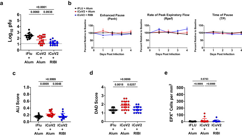
Fig. 5. Vaccine immune serum promotes cross-protection with modest pathology during heterologous infection.
a Pulmonary viral titers at 5 days post-infection (DPI) following Rs-SHC014-CoV infection in naïve mice that received a passive serum transfer from mice vaccinated with inactivated influenza virus adjuvanted with aluminum hydroxide (iFLU + Alum, n = 18), inactivated SARS-CoV-2 adjuvanted with aluminum hydroxide (iCoV2 + Alum, n = 19), or inactivated SARS-CoV-2 adjuvanted with Sigma Adjuvant System adjuvant (iCoV2 + RIBI, n = 14); dotted line represents assay limit of detection; pfu plaque forming units. b Pulmonary function measured by whole-body plethysmography following challenge with Rs-SHC014-CoV in naïve mice that received a passive serum transfer from mice vaccinated with iFLU + Alum (n = 6), iCoV2 + Alum (n = 6), or iCoV2 + RIBI (n = 6); results reported as group mean ± standard error of the mean; *p < 0.05, iFLU + Alum versus iCoV2 + Alum, two-way ANOVA with Geisser-Greenhouse correction and Tukey’s multiple comparisons correction. c, d Acute lung injury (ALI) (c) and diffuse alveolar damage (DAD) (d) scores in hematoxylin and eosin (H&E)-stained lungs at 5 DPI following Rs-SHC014-CoV infection in naïve mice that received a passive serum transfer from mice vaccinated with iFLU + Alum (n = 18), iCoV2 + Alum (n = 19), or iCoV2 + RIBI (n = 14). e Quantification of pulmonary eosinophils immunohistochemically labeled for eosinophil peroxidase (EPX, brown cells) at 5 DPI following Rs-SHC014-CoV infection in naïve mice that received a passive serum transfer from mice vaccinated with iFLU + Alum (n = 18), iCoV2 + Alum (n = 19), or iCoV2 + RIBI (n = 13). Individual data points represent independent biological replicates; solid lines and error bars represent group mean ± standard deviation; data analyzed by Kruskal–Wallis test with Dunn’s multiple comparisons correction; solid horizontal lines above data represent pairwise comparisons with p values; data presented as combined results from one (b) or two (a, c, d, e) independent animal experiments.

