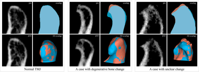
Figure 1.
Representative images of temporomandibular joint (TMJ) cone-beam computed tomography (CBCT) images and image processing result. Sagittal CBCT images of pre-treatment (upper row) and post-treatment (lower row) condyle from a normal subject and patients with degenerative joint diseases. Overlap visualizes the comparison of segments between pre- and post-treatment condylar heads. Red segment, pre-treatment; Blue segment, post-treatment. 3D overlap shows rendered models of pre- and post-treatment condyle. Red segment, pre-treatment; Blue segment, post-treatment. The images were generated using the open-source software 3D Slicer (v4.11, Slicer, http://www.slicer.org/).

