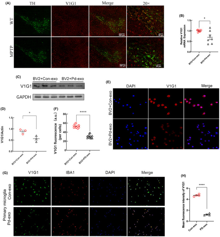
FIGURE 3.

Lysosomal VIG1 was reduced in MPTP‐induced PD models and PD‐exo‐treated BV2 cells. (A) Representative images of double immunolabeling analysis for lysosomal V1G1 with either dopaminergic neuron marker TH in the SN of MPTP‐treated mice. (B) The mRNA levels of lysosomal V1G1 were detected by RT‐PCR in the PD‐exo‐treated microglia or Con‐exo‐treated microglia. (C, D) The lysosomal V1G1 was detected by western blot after the microglia were treated with PD‐exo or Con‐exo. The protein levels were normalized to β‐Actin. (E, F) Representative immunostaining and quantification of lysosomal V1G1 in the PD‐exo‐treated BV2 cells or Con‐exo‐treated BV2 cells. (G, H) Representative immunostaining and quantification of lysosomal V1G1 in the PD‐exo‐treated primary microglia or Con‐exo‐treated primary microglia. *p < 0.05, ****p < 0.0001.
