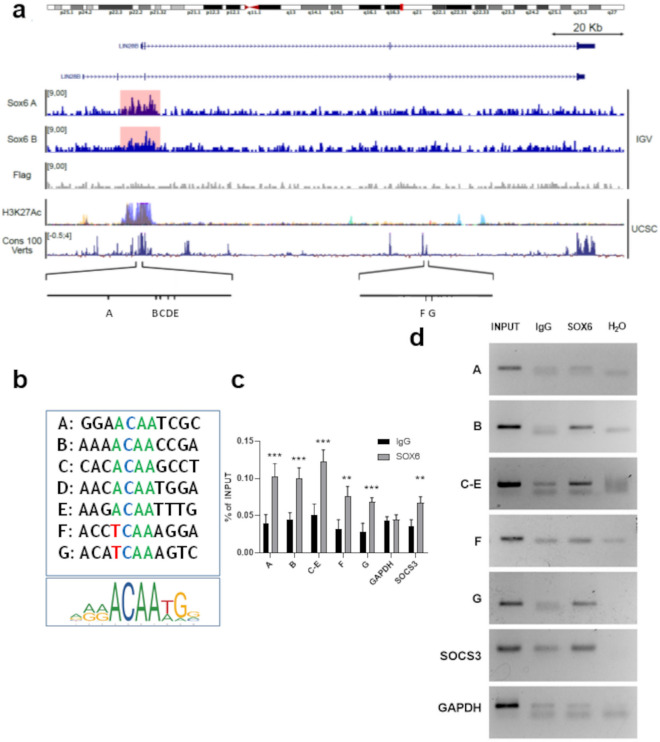
Figure 5.
Identification of the SOX6 binding sites within the LIN28B locus. (a) Lin28B genomic locus visualized in Integrative Genomic Viewer (IGV) and with the UCSC genome browser (https://genome.ucsc.edu/). From the top: chromosome representation (IGV), gene transcripts (UCSC), two SOX6 CUT&RUN replicates and the respective negative Flag control in HUDEP1 cells (IGV), H3K27ac overlap (UCSC), vertebrate conservation (UCSC) and positions of the SOX6 binding sites within the promoter (A–E) and the third intron (F–G). (b) SOX6 sites compared with the SOX6 consensus Jaspar matrix 515.1. (c) Chromatin IP results obtained in HEL cells expressing SOX6. GAPDH locus: negative control; SOCS3 promoter: positive control34. Histograms represent the results of three biological replicates (n = 3, each of them in three technical replicates, as analyzed by RTqPCR (Error bars: standard error of mean; *p < 0.05; **p < 0.01; ***p < 0.001). (d) representative ChIP result. Antibodies are listed in the Supplementary table 2.

