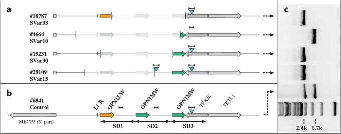
Fig. 1. Presence of the SDIns in BCM patients with partial deletions of the OPN1LW-OPN1MW gene cluster.
Schematic representation of the OPN1LW-OPN1MW gene cluster in patients with partial deletions with the extent of the deletions marked by brackets (a) in comparison with a normal control (b) with one OPN1LW gene (red arrow) and two OPN1MW gene copies (green arrows). SD1, SD2 and SD3 indicate the extent of individual segmental duplications (SD) forming the opsin gene cluster. The blue triangles indicate the presence and localization of the 697 bp SDIns. The long distance PCR amplicon LD-SDIns covering the region of SDIns and flanking sequences which was used to distinguish between presence (2.4 kb PCR product) or absence (1.7 kb PCR product) of the SDIns is indicated by dumbbell-shaped marks. The agarose gel separation of the long distance PCR products for the patients with deletions and the control is montaged alongside (c) including a 1 kb DNA ladder size standard at the bottom. Note that the presence of two copies of the SDIns in proband #28109 was further supported by qPCR.

