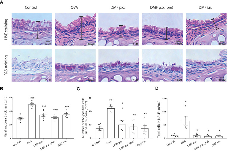
Figure 3.
DMF reduced the infiltration of nasal mucus and NALF in the allergic asthma model. (A) The histology of nasal mucosa in treatment groups has a less epithelium thickness of nasal mucosa and accumulation of infiltrated inflammatory cells, inflammatory response, and goblet cells. Representative H&E or PAS-stained images of nasal mucosa were shown. Scale bar=10uM. (B, C) the bar graphs show the nasal mucosa thickness (B) and the PAS-positive cells (C) in the nasal mucosa in OVA-induced DMF-treated mice. (D) The total cells were calculated from the NALF collected from the in vivo experimental groups. Results are shown as the Mean ± SEM (n = 5-6 per group). #P < 0.05, ##P < 0.01, ### < 0.001 versus the control group. ***P < 0.001, **P < 0.01, *P < 0.05 vs OVA group. NALF, nasal lavage fluid.

