Figure 7.
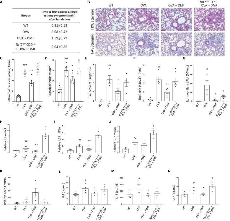
Oral administration of DMF alleviates the inflammatory response in the lungs of asthmatic mice. (A) The allergic symptoms, such as rubbing, sneezing, and abdominal respiration, were monitored for their first appearance in OVA-induced WT or Nrf2fl/flCD4cre mice after p.o. administration of DMF. (B–E) Histological analysis was performed on the lung tissue collected from the OVA-induced WT or Nrf2fl/flCD4cre mice after p.o. DMF treatment using H&E and PAS staining. Inflammation score, bronchial thickness, and PAS staining score were calculated. (F, G) After DMF treatment, the total number of immune cells and eosinophils was calculated through the microscopic examination of BALF collected from the OVA-induced WT or Nrf2fl/flCD4cre mice. (H, I) RT-qPCR was performed to examine the expression of Th2 cytokines IL4 (H), IL13 (I), and Th17 cytokine IL17 (J) in the lung of OVA-induced WT or Nrf2fl/flCD4cre mice in the presence or absence of DMF. (K) RT-qPCR was utilized to study the Treg transcription factor Foxp3 (K) level in the lung of OVA-induced WT or Nrf2fl/flCD4cre mice in the presence or absence of DMF. (L–N) ELISA was conducted to inspect the level of circulatory Th2 cytokines IL4 (L), IL13 (M), and Th17 cytokine IL17 (N) in the lungs of OVA-induced WT or Nrf2fl/flCD4cre mice in the presence or absence of DMF. Time in (A) is shown as average ± SD (min). All other data are shown as the mean ± SEM (n = 6 per group). #P < 0.05, ##P < 0.01, ###P < 0.001 versus the WT group. ***P < 0.001, **P < 0.01, *P < 0.05 vs OVA group or OVA+DMF group. Pa < 0.05, Pb < 0.01 and Pc < 0.001.

