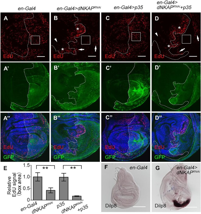
Figure 4.
Aberrant cell proliferation in dNKAP-knockdown wing discs. (A–D’’) Decreased EdU signal in a population of dNKAP-depleted cells. Boxes indicate the pouch region. Note that EdU signals are increased in a patch of dNKAP-depleted cells (indicated by arrows) and the adjacent wild-type cells (indicated by stars) along the anterior–posterior boundary but decreased in the wild-type cells far from dNKAP-depleted cells in the anterior compartment (indicated by arrowheads). GFP was used to mark the knockdown domain. Shown are single confocal sections of third-instar wing imaginal discs with posterior side to the right and dorsal side up. Scale bar, 50 μm. (E) Quantification of EdU signals in the boxed area of the wing disc. n = 3. **P < 0.01. (F and G) Increased Dilp8 mRNA level in dNKAP-knockdown wing discs. Scale bar, 100 μm.

