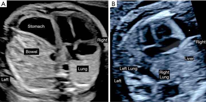
Figure 4.
Ultrasound appearance of left and right diaphragmatic hernia in the second trimester. Prenatal ultrasound images obtained in a patient with left diaphragmatic hernia at 26 weeks’ gestation (A) and a right diaphragmatic hernia at 22 weeks’ gestation (B). The left diaphragmatic hernia is characterized by a right mediastinal shift of the heart and intrathoracic bowel and stomach herniation. The contralateral right lung is posterior to the right atrium and there is reduction of the observed to estimated lung to head ratio to a moderate hernia severity. In right diaphragmatic hernia the cardiac axis is rotated to the left and the mediastinal shift is less marked despite intrathoracic herniation of the liver. Both lungs measure smaller in size and there is a pericardial effusion (*) which is common in this setting.

