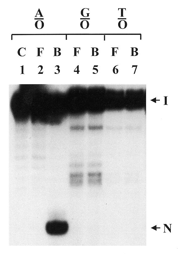
Figure 5.

Analysis of DNA cleavage in mtMYH-bound DNA fractions. Calf mtMYH (210 µg) was incubated in the standard gel mobility assay for 60 min at 37°C with 30 fmol 3′-end labeled 20mer duplex DNA and 200 ng of poly(dI-dC). After analysis on an 8% polyacrylamide gel, the enzyme-bound and enzyme-free DNA bands were excised from the gel and electroeluted. The fractions with equal amount of radioactivity in the loading dye were heated at 90°C for 2 min and fractionated on a 14% polyacrylamide–7 M urea sequencing gel. The mismatches used were A/8-oxoG (A/O), G/8-oxoG (G/O) and T/8-oxoG (T/O). DNA with A/8-oxoG was run on the control lane (C). F and B represent samples from enzyme-free and enzyme-bound DNA bands, respectively. Arrows indicate the intact DNA substrate (I) and the cleaved DNA fragment (N).
