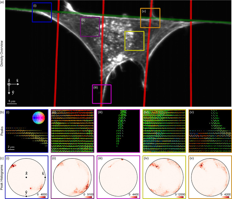
Fig. 5. pol-diSPIM measurements of phalloidin-labelled 3T3 mouse fibroblasts grown on nanowires show dipoles oriented parallel to their nearest nanowires and reveal distinct out-of-plane dipole populations across the cell.
(a) Reconstructed density maximum intensity projection of a cell grown on crossed nanowires, with hand-annotated wires measured from a wirespecific channel highlighted with red and green lines. ROIs (i-iii) are outlined in color and examined in subsequent panels. (b) Peak cylinders drawn every 780 nm in regions with total counts > 5000, colored by orientation (see inset color hemisphere), with lengths proportional to the maximum diameter of the corresponding ODF. (c) Histogram of all peak cylinders with total counts > 5000 in each ROI. Bins near the edge of the circle indicate in-plane orientations, bins near the center indicate out-of-plane orientations, and dots mark the Cartesian axes on the histogram.

