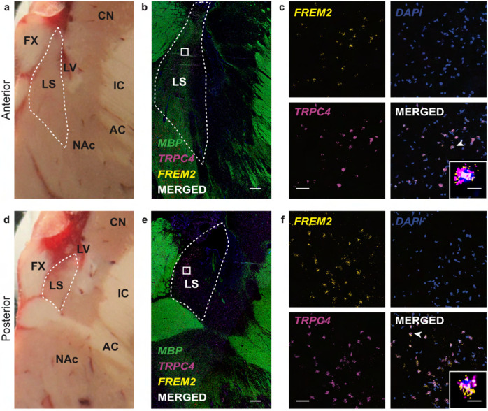
Figure 4. In situ validation of FREM2 expression in the lateral septum (LS).
a, d, Postmortem human brain block from a donor not include in the snRNA-seq study shown at the level of anterior LS (a) and posterior LS (d). LS is outlined by the dashed white line. CN – caudate nucleus; FX – fornix; LS – internal capsule; AC – anterior commissure; NAc – nucleus accumbens. b, e, 2x magnification smFISH images at the anterior LS (b) and posterior LS (e) illustrating expression of TRPC4 and FREM2. MBP expression is included for anatomical visualization of white matter. LS is outlined by the dashed white line. White squares indicate the approximate location of the corresponding 40x images. White bar indicates 1000μm. c, f, 40x magnification smFISH images of the area demarcated by white squares in 2x images from anterior LS (c) and posterior LS (f). White bar indicates 50μm. Merged images shows TRPC4 and FREM2 co-expressing cells in white. Inset depicts a zoomed in image of a single neuron indicated by the white arrowhead. White scale bar of the inset indicates 10μm.

