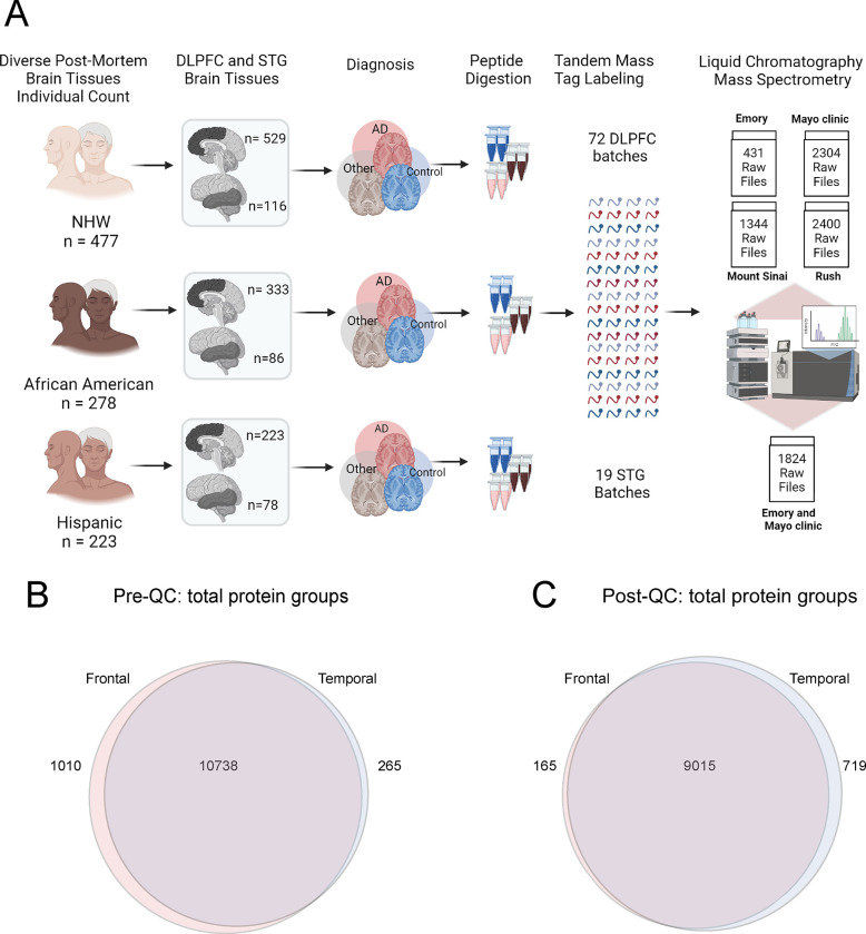
Fig 1. A. Schematic illustrating the cohort characteristics and the experimental workflow for mass spectrometry (MS) of the human brain proteome across frontal and temporal brain tissue samples.
This study incorporated a total of 1105 dorsolateral prefrontal cortex (DLPFC) brain tissues from 998 individuals, categorized as follows: 529 non-Hispanic white (NHW), 333 African American, 223 Latino American, and others (n= 20) as applicable. These samples were sourced from four prominent data distribution sites: Emory University, Mayo Clinic, Rush University, and Mount Sinai University Hospital. Additionally, 280 STG tissues from a subset of 244 individuals were included, with 116 NHW, 86 African American, 78 Hispanic, and others as applicable. STG samples were obtained from a racially diverse set of specimens originating from Mayo Clinic and Emory, distributed across 19 batches. Tissues underwent an experimental pipeline involving protein digestion, batch randomization, TMT labeling, fractionation, and subsequent mass spectrometric measurements. A total of 72 DLPFC batches were processed, comprising 9 batches from Emory, 24 from Mayo Clinic, 14 from Mount Sinai, and 25 from Rush (comprising a total of 72 batches). The randomization of batches was conducted to ensure a representative and diverse dataset. The output included a total of 6479 raw files for DLPFC samples and 1824 raw files for STG. B. Venn diagram of total number of proteins quantified from DLPFC and STG samples. A total of 11748 protein groups were identified from DLPFC and 11003 from STG samples, with 10738 shared protein groups. C. Venn diagram of total protein from DLPFC and STG samples after quality control (QC) across all samples. 9180 protein groups were identified from DLPFC samples and 9734 from STG, with 9015 shared protein groups.

