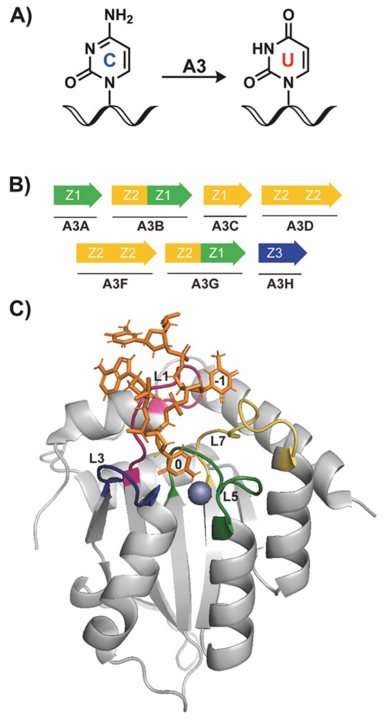
Figure 1.

Overview of APOBEC3 protein structure and function. A) A3 enzymes deaminate at target cytosine (C) bases in ssDNA, converting the nucleobase to uracil (U). B) The seven members of the A3 family are either single domain (A3A/C/H) or double domain (A3B/D/F/G) enzymes. Colors represent different phylogenetic grouping, based on domain sequence. C) Structure of A3Bctd bound to a single-stranded 5-mer oligonucleotide (orange). In this structure, the 0 on the oligonucleotide indicates the target cytosine which is deaminated by A3 proteins, and −1 indicates the 5’ nucleotide, which makes extensive interactions with the protein to impart binding selectivity. Highlighted loops are loops 1 (L1, pink), 3 (L3, blue), 5 (L5, green), and 7 (L7, yellow), which are also important in determining substrate selectivity. The blue sphere is a coordinated zinc ion in the active site, which is required for enzymatic activity. PDB: 5TD5.
