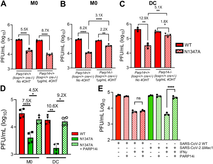
Fig. 4. PARP14 inhibits the replication of Mac1-mutant MHV and SARS-CoV-2.
A) Parp14 floxed cre+/− BMDMs were treated with and without 4-OHT. 24 hours later these cells were infected with WT and N1347A MHV at an MOI of 0.1. At 20 hpi cells and supernatants were collected and progeny virus was quantified by plaque assay. B-C) PARP14 floxed cre+ BMDMs (B) and DCs (C) were treated with or without 4-OHT. 24 hours later these cells were infected with WT and N1347A MHV at an MOI of 0.1. At 20 hpi cells and supernatants were collected and progeny virus was quantified by plaque assay. Data shown in A-C are from 1 experiment and are representative of 3 independent experiments with N=3 for each experiment. D) PARP14+/+ BMDMs and DCs were infected with WT and N1347A MHV at an MOI of 0.1 and then treated with DMSO or PARP14i (1 μM). At 20hpi cells and supernatants were collected and progeny virus was quantified by plaque assay. Data shown in D are from 1 experiment and are representative of 3 independent experiments with N=4 for each experiment. E) WT Calu-3 cells were either mock-treated or IFNγ-treated (50 Units) O/N. These cells were then infected with WT and ΔMac1 SARS-COV-2 at an MOI of 0.1 and treated with DMSO or PARP14i (1 μM) at 1 hpi. At 48 hpi cells and supernatants were collected and progeny virus was quantified by plaque assay. Data shown in E are from 1 experiment and are representative of 3 independent experiments with N=3 for each experiment.

