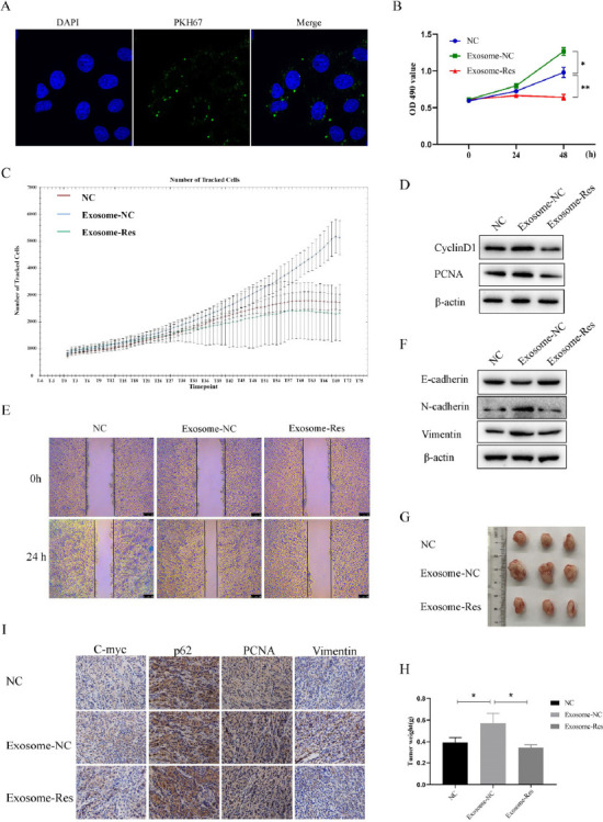
Fig. (4).

Resveratrol-induced exosomes inhibited the progression of HCC. (A) Absorption of exosomes stained by PKH was measured by high-resolution laser confocal fluorescence microscopy. (B and C) Cell viability and proliferation of Huh7 cells treated with Exosome-NC or Exosome-Res were measured by MTT and high-content cell imaging analyzer, respectively. (D) The expression of cyclinD1 and PCNA was detected by Western blot. (E) Wound healing assay was performed to evaluate the cell migration. (F) The expression of EMT markers was detected by Western blot. (G) The tumor extracted from the xenograft mice treated with different exosomes. (H) The tumor weight was measured in different groups. I, The expression of c-myc, p62, PCNA and vimentin in tumors were determined by immunohistochemistry.
