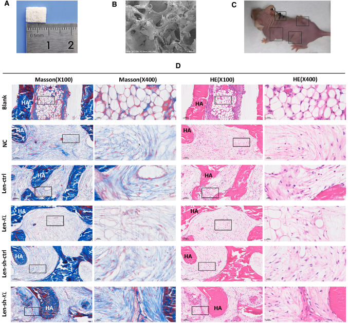
Fig. 4.
Hematoxylin and eosin (HE) staining and Masson's trichrome staining for subcutaneously transplanted hydroxyapatite (HA) scaffold materials containing human renal interstitial fibroblasts (hRIFs) in nude mice. A A porous HA scaffold material with a size of 8.5 mm × 5 mm × 1.5 mm. B The porous structure of HA, as visualized by scanning electron microscopy. C Representative picture of a 16-week-old female nude mouse in which HA scaffold materials containing hRIFs were subcutaneously transplanted into 4 sites on the dorsal surface. D HE and Masson staining of the regenerated collagen tissue in grafts. There were 6 groups: the blank group (HA without cells; n = 6), the normal control (NC) group (HA with hRIFs; n = 6), the Len-sh-ctrl group (HA with hRIFs transfected with Len-sh-ctrl; n = 6), the Len-sh-KL group (HA with hRIFs transfected with Len-sh-KL; n = 6), the Len- ctrl group (HA with hRIFs transfected with Len-ctrl; n = 6) and the Len-KL group (HA with hRIFs transfected with Len-KL; n = 6)

