Fig. 5.
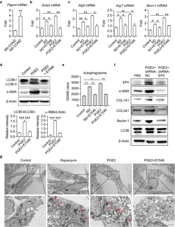
PGE2-induced autophagy in HSCs was restrained by EP4 inhibitor E7046. a The mRNA level of EP4 coding gene Ptger4 in LX-2 cells. LX-2 cells were cultured with M0- or M2-conditional medium (COND) for 48 h and total RNA was isolated for real-time PCR assay. b, c The mRNA levels of Acta2, Atg5, Atg7 and Becn1 in LX-2 cells were detected by real-time PCR. LX-2 cells were cultured with M2-conditional medium or 100 ng/mL PGE2, in the presence or absence of 1 μM E7046 for 48 h. d The expression levels of LC3B-I, LC3B-II and α-SMA in LX-2 cells were determined by Western blot. Cells were cultured with 100 ng/mL PGE2, in the presence or absence of 1 μM E7046 for 48 h. The intensity of α-SMA blot was quantified with ImageJ software. The conversion ratio of LC3B-I to LC3B-II was calculated and the expression level of α-SMA was normalized to β-Actin. e Autophagosome in LX-2 cells. Cells were stained with autophagy-specific dye and analyzed by microplate reader. f The expression levels of EP4, α-SMA, COL1A1, COL3A1, Beclin-1, and LC3B in LX-2 cells were detected by Western blot. LX-2 cells were transfected with EP4 shRNA and stimulated with recombinant PGE2. g The bilayer autophagosomes and lysosomes were observed with transmission electron microscope. Scale bar = 5 μm (top panel). The panels on the bottom are higher-magnification images of the cropped regions. Scale bar = 1 μm (bottom panel). P-values were obtained by unpaired t test (two groups) or one-way ANOVA (multiple groups). *P < 0.05, **P < 0.01 and ***P < 0.001 compared with controls

