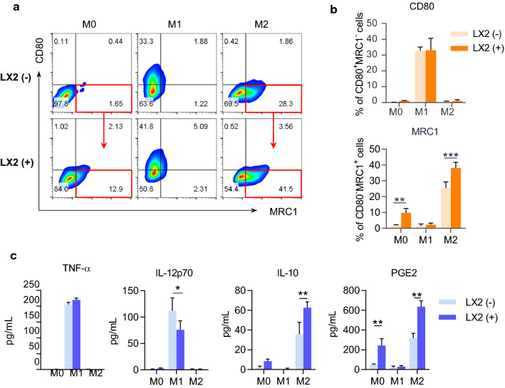
Fig. 8.
LX-2 cells promoted M2 macrophage polarization and PGE2 secretion. BMDMs were cultured in the presence or absence of LX-2 cells for 48 h. BMDMs and the cell culture supernatant were collected to identify cell polarization phenotypes. a Representative flow cytometry plots displaying the expression of CD80 (M1 marker) and MRC1 (M2 marker) gated on F4/80+ BMDMs. b Percentage of CD80+MRC1− (M1) and CD80−MRC1+ (M2) subsets in BMDMs. c The cytokine concentrations in the culture media of BMDMs. Data represent mean ± SD (n = 4 in each group). P-values were obtained by Student t test or Mann–Whitney test. *P < 0.05, **P < 0.01 and ***P < 0.001 between indicated groups

