Fig. 8.
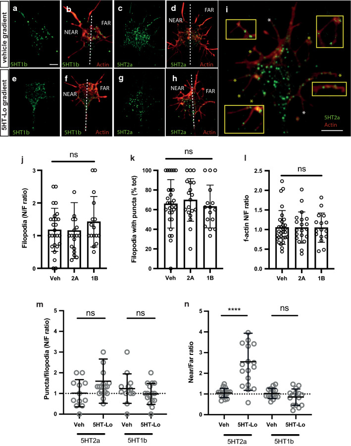
5HT-Lo exposure causes asymmetric distribution of membrane 5-HT2a receptors to the motile side of growth cones. a–h Representative growth cones exposed to vehicle and 5HT-Lo gradients (micropipette on upper left side) and stained for membrane localization of receptors 5-HT1b (green, a, e), 5-HT2a (green, c, g) and merged with actin, (red, b, d, f, h). Dotted line separates the near and far regions of the growth cone. i Increase magnification of representative image h to show the presence of 5-HT2a receptor membrane on most filopodia (*) of the growth cone (yellow *, insets). Scale bars are 5 μm. (j-k)There was no significant near/far bias in the amount of f-actin (/area) (j) and the number of filopodia (k) in growth cones exposed to vehicle and 5HT-Lo. Kruskal–Wallis, Dunn’s multiple comparison post hoc test. (l) There was no significant bias in the total percentage of filopodia containing 5HT2a and 5HT1b puncta (60%). One-way ANOVA followed by Tukey’s multiple comparison post hoc test. (m) There was no significant near/far bias in the amount of 5-HT2a and 5-HT1b puncta per filopodia. (n) Analysis of 5-HT receptor membrane localization showed translocation of 5-HT2a (n = 18, p = <0.0001) to the near side of growth cones exposed to 5HT-Lo while no significant translocation of 5-HT1b (n = 15, p = 0.346) was observed. (Mann–Whitney U-test)

