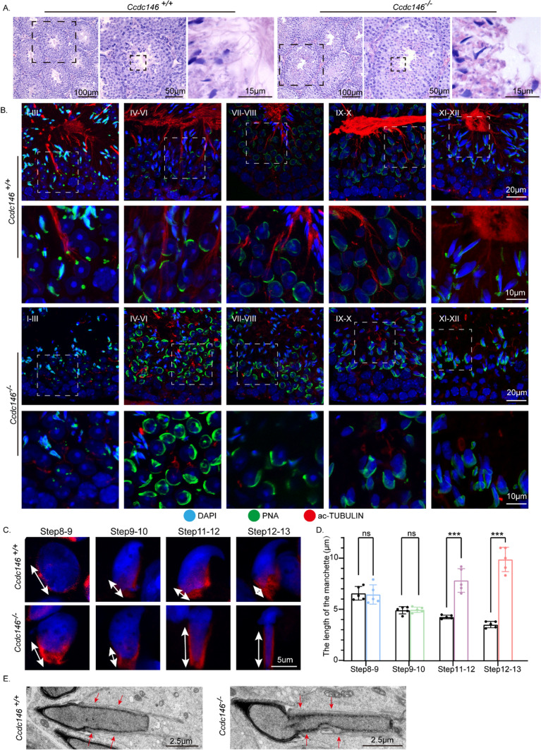
Fig. 4.
Disorganized flagellum and manchette in Ccdc146−/− spermatids. A The histology of the seminiferous tubules from Ccdc146+/+ and Ccdc146−/− male mice. B Immunofluorescence analysis of Ac-TUBULIN (red) and PNA lectin (green) to identify sperm flagellum biogenesis. C Abnormal manchette elongation in Ccdc146−/− spermatids. Spermatids from different manchette-containing steps were stained with α/β-TUBULIN antibody (red) to visualize the manchette. Ccdc146−/− spermatids displayed abnormal elongation of the manchette. D Statistical analysis of the length of the manchette. The color columns indicate the manchette length of the Ccdc146−/− spermatids in different steps, while the black columns indicate the manchette length of the Ccdc146+/+ spermatids. Data are presented as means ± SEM. two-tailed Student’s t test; ns no significance; ***P < 0.001. E TEM revealed that the manchette of elongating spermatids (steps 9–11) from Ccdc146.−/− mice were ectopically placed. The red arrowhead indicates the abnormal manchette. Scale bars: 100 μm (A); 50 μm (A); 20 μm (B); 15 μm (A); 10 μm (B); 5 μm (C); 2·5 μm (E)

