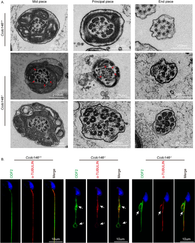
Fig. 5.
Impaired ODF transportation in Ccdc146−/− spermatids. A Cross-sections of Ccdc146−/− sperm tails revealed the disorganization of axonemal microtubules and tail accessory structures. Red arrowheads indicate an increase in outer dense fibers. B Immunofluorescence of ODF2 (green) and α-TUBULIN (red) in spermatids from Ccdc146+/+ and Ccdc146−/− mice. Nuclei were stained with DAPI (blue). Scale bars: 250 nm (A); 10 μm (B)

