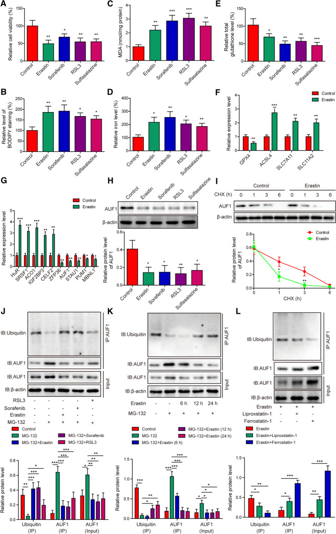
Fig. 1.
Ubiquitin–proteasome pathway mediates AUF1 degradation in AECs during ferroptosis. AECs were treated with Erastin (5 μM), Sorafenib (10 μM), RSL3 (10 μM), or Sulfasalazine (20 μM) for 12 h. Ferroptosis was assessed by measuring cell viability using MTT assay (A), BODIPY staining (B), intracellular MDA (C), iron (D), and total glutathione E levels. F The expression levels of ferroptosis-related markers GPX4, ACSL4, SLC7A11, and SLC11A2 were examined by RT-PCR in control or Erastin-treated AECs. G The expression levels of indicated RBPs were examined by RT-PCR in control or Erastin-treated AECs. H The protein level of AUF1 was examined by western blot in AECs stressed with Erastin, Sorafenib, RSL3, or Sulfasalzine. I The degradation of AUF1 protein was examined by CHX chase assay, where AECs were treated with CHX (5 μg/mL) for indicated time periods. The ubiquitination of AUF1 was examined by Co-IP using anti-AUF1 antibody, after proteasome signaling of AECs was blocked with MG-132 (25 μM) for 2 h, then AECs were treated with Erastin (5 μM), Sorafenib (10 μM), or RSL3 (10 μM) for 12 h (J); after proteasome signaling of AECs was blocked with MG-132 (25 μM) for 2 h, then AECs were treated with Erastin (5 μM) for indicated time periods (K); after AECs were treated with Erastin (5 μM), without or with Liproxastatin-1 (25 nM) or Ferrostatin-1 (100 nM) for 12 h (L). *P < 0.05, **P < 0.01 and ***P < 0.001

