Fig. 5.
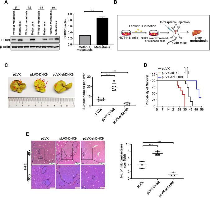
DHX9 facilitates liver metastasis of colorectal cancer cells. A Western blotting analysis of DHX9 in primary CRC tumors from non-metastatic and liver metastatic patients and quantitative data of DHX9 protein levels were shown Data were represented as mean ± SEM (n = 3). **P < 0.01; Student’s t test. B Graphic illustration for liver metastasis mouse model of CRC. C Representative images of metastatic liver in vector (pLVX), pLVX-DHX9 and pLVX-shDHX9 group were shown (left) and surface nodules were counted (right). Arrows indicated metastatic foci on liver surface. n = 6 mice per condition. Data were represented as mean ± SEM. ***P < 0.001, one-way ANOVA with post hoc intergroup comparison by Tukey's test. D Survival curves analyzed with Kaplan–Meier in the indicated groups of mice were shown (n = 6). Log-rank test, ***P < 0.001. E H&E staining of liver sections, dot plots indicated metastatic nodules. Left, representative images of metastatic nodules in H&E-stained liver section were shown. Scale bar, 500 µm (40 ×), 200 µm (100 ×). Right, quantification of liver metastatic nodules in microscopic fields of 40 × was shown. n = 3 mice per condition. Data were represented as mean ± SEM. ***P < 0.001, one-way ANOVA with post hoc intergroup comparison by Tukey's test

