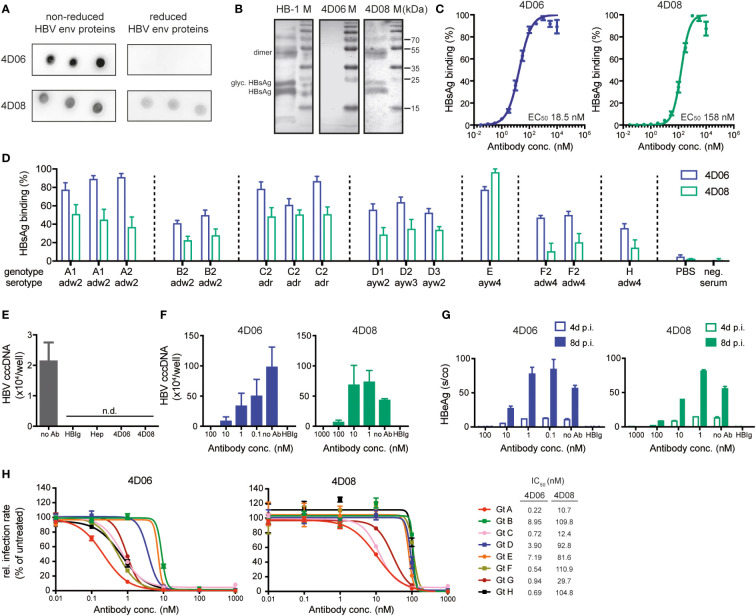
Figure 2.
Characterization of mAbs 4D06 and 4D08. (A) Dot blot analysis of the interaction between mAbs and HBV envelope (HBV env) proteins blotted on a PVDF membrane under reducing or non-reducing conditions. 4D06 and 4D08 served as primary antibodies, goat anti-human IgG HRP was used for detection. (B) Western blot analysis of the interaction of HBV env proteins with mAbs. HBV env proteins were separated by SDS PAGE under reducing conditions and blotted on a PVDF membrane. Env protein detection was performed as described in (A). Bands represent non-glycosylated and glycosylated (glyc.) forms of monomeric HBsAg and dimeric (dimer) HBsAg. HB-1 served as a positive control for an antibody recognizing a linear epitope. (C) ELISA analysis of the interaction between mAbs and plate-bound HBsAg; polyclonal goat anti-human IgG HRP antibody was used for detection. EC50 values were calculated by nonlinear regression. (D) ELISA analysis for the interaction between mAbs and HBsAg from several HBV geno- and serotypes, based on 15 HBsAg-positive human plasma samples from the Paul-Ehrlich Institute (Langen, Germany). HBsAg was captured with HBsAg-specific antibodies from a commercial anti-HBs ELISA kit (DiaSorin, Italy). The binding of biotinylated mAbs 4D06 and 4D08 was detected with Avidin-HRP. Data points are normalized to the highest value. (E) HBV uptake assay with differentiated HepG2-NTCP cells inoculated with HBV after pre-incubation with heparin (Hep), HBIg (0.3 IU/well), 4D06 or 4D08 (100 nM). No Ab served as neg. control. Absolute quantification of intracellular HBV cccDNA 24 hours post-infection (p.i.). n.d. = not detectable. (F) Neutralization assay: Antibodies were pre-incubated with HBV, followed by inoculation of HepG2-NTCP cells with HBV. Absolute quantification of intracellular HBV cccDNA eight days p.i.; IC50 determination with non-linear regression. (G) Level of HBeAg in the cell culture supernatant four and eight days p.i. (H) Huh7-NTCP cells were preincubated with 4D06 or 4D08 and inoculated with HDV, enveloped with HBV proteins of genotypes (Gt) A-H. Cells were analyzed for HDAg by immunofluorescence assay 7 days p.i. IC50 concentration of 4D06 and 4D08 were calculated with non-linear regression. All data points represent mean values ± SD from triplicates.

