Fig. 1.
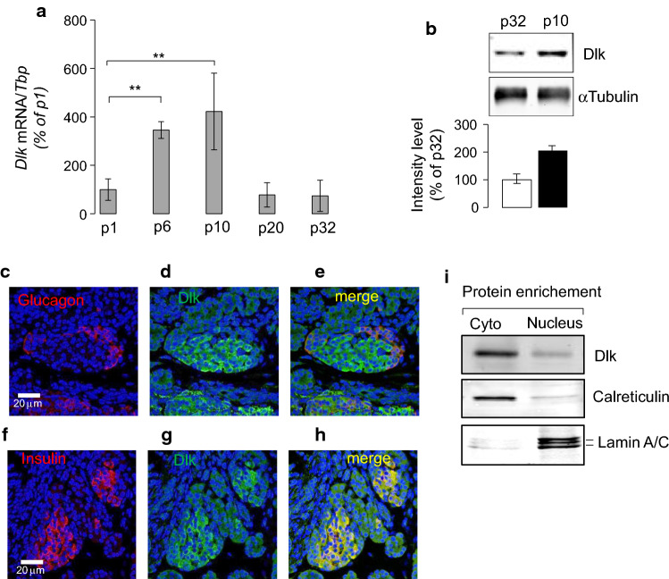
Postnatal islet induction and beta-cell nuclear localization of Map3k12 (Dlk) in neonate rats. a The expression of the Map3k12 (Dlk) was measured by a RT-qPCR and b Western blotting in isolated islet of newborn rats between p1, p6, p10 and p20 postnatal days and young adult rats (p32). The mRNA levels of Map3k12 (Dlk) were normalized to those of Tata box binding protein (Tbp) and were expressed as % changes. The expression of Map3k12 (Dlk) at p1 was set to 100%. The data are the mean ± SEM of 3 independent experiments performed in triplicate (**p < 0.01, by ANOVA). b Western blotting was performed using total proteins (20 μg/lane) isolated from islets of 10- and 32-day-old rats using an anti-Dlk (1/1000; Genetex) and anti-αTubulin (1/5000; Sigma) antibodies. The intensities of the bands were quantified using ImageJ, taking care to avoid saturation and subtracting the background. Values were expressed as the integrals (area × mean density) of each band (normalized to αTubulin band). Values from p32 were set to 100%. c–h Representative immunofluorescence images for Map3k12 (Dlk), insulin and glucagon in pancreatic islets of 10-day-old rat (×40 magnification) using anti-Dlk (1/1000; Genetex), anti-insulin (1/100, Dako) and anti-glucagon (1/100, Sigma) antibodies. The scale bars in each picture row correspond to 20 μm. c Map3k12 (Dlk) (green; d, g), and insulin (red; f, h) performed on fixed pancreas of newborn rats of 10 days old. Merged images were achieved to show the potential co-localization of Map3k12 (Dlk) with glucagon (e) or insulin (h). Yellow indicates the co-localization. Blue, DAPI. i Detection of Map3k12 (Dlk) protein in islets nuclear-enriched fraction of 10-day-old rats. Nuclear and cytosol (Cyto) proteins (20 μg/lane) were loaded onto 10% SDS-PAGE and then detected by Western blotting using an anti-Dlk (1/1000; Genetex), anti-Lamin a/c (1/1000; Abcam) and anti-Calreticulin (1/1000; Abcam) antibodies

