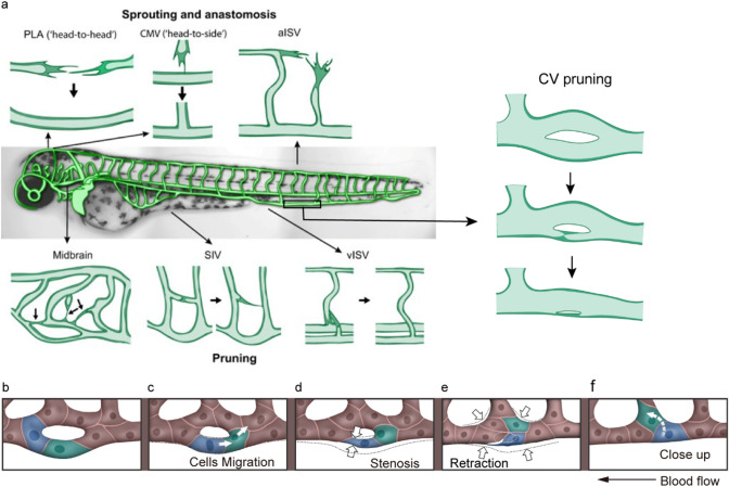
Fig. 3.
Anastomosis and vessel pruning events in the zebrafish model. a The process of anastomosis and vessel pruning in zebrafish vasculature. The central panel shows an overview of the zebrafish vascular beds. Sprouting and anastomosis have been studied in the palatocerebral artery (PLA), the communicating vessel (CMV) and the segmental arteries (aISV). Vessel pruning has been studied in the midbrain vasculature, the subintestinal vein (SIV), the segmental veins (vISV) and the caudal vein (CV). Figure a adapted from Charles et al. [34]. The process of zebrafish CV pruning is mediated by EC rearrangement, which includes the stages of selection pruning segment (b), cell migration (c), stenosis (d), retraction (e) and close-up (f). b The diameters of the lower branch and the upper branch are the same at the beginning of vessel pruning. c, d The ECs marked with blue and green migrate against the blood flow, resulting in vessel stenosis at the lower branch. e, f The ECs marked with blue and green migrate into the adjacent vessel, finishing the pruning of the lower branch. The arrow indicates the direction of blood flow. The rearrangement of ECs during vessel pruning is marked with blue and green. The arrows in (c, f) indicate the direction of EC migration. The arrows in (d, e) indicate vessel stenosis. Figure b–f adapted from Wen et al. [28]

