Fig. 1.
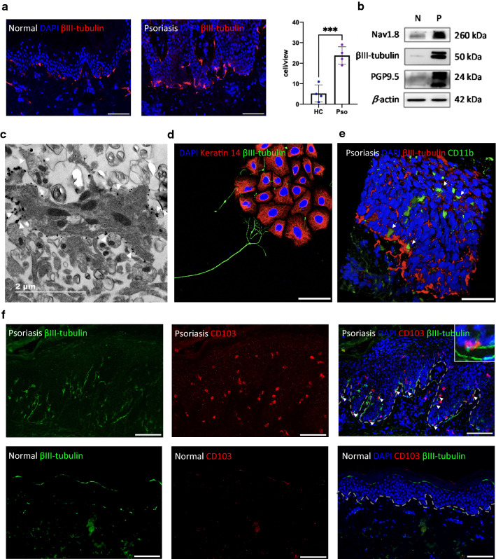
Location and expression of nerves in psoriasis lesions. a IF of βIII-tubulin in healthy and psoriatic lesional skin (left). Qualification of innervated cells (right), scale bar = 100 μm. b Western blotting of βIII-tubulin, Nav1.8, PGP9.5 and β-actin in the epidermis of normal skin (N) and psoriatic lesion (P). c Immunoelectron microscopy to detect βIII-tubulin in psoriatic epidermis, scale bar = 2 μm. d IF to detect the localization of mouse keratinocytes (K14, red) and DRG neurons (βIII-tubulin, green), scale bar = 50 μm. e IF of CD11b (green) and βIII-tubulin (red) in psoriatic lesion, white arrow marked CD11b+ cells, scale bar = 50 μm. f IF of CD103 (red) and βIII-tubulin (green) in psoriatic lesion, white arrow marked CD103+ cells co-localized with nerve fibers, scale bar = 100 μm

