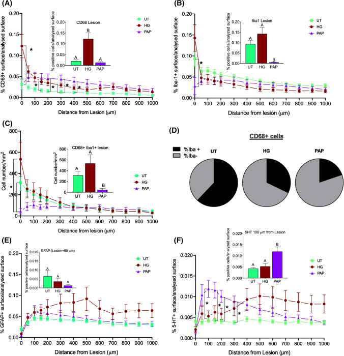
Fig. 6.
Papilla implantation significantly reduce the number of activated macrophages and increase the percentage of 5-HT positive cells in the spinal cord lesion Papillae (PAP) were implanted in a rat spinal cord hemisection model (controls were fibrin hydrogel (HG) and untreated hemisection (UT)) for one week. Then, spinal cord tissue centered on the lesion (1 cm in each direction) was processed for immunofluorescence. Staining was quantified at the lesion (histograms inserted in A, B, C, D and F) and away from the lesion in concentric circles up to 1 mm (A, B, C, D and F) (n = 3). Stained area for activated microglia: CD68 A and Iba1 B, C Number of cells positive for both CD68 and Iba1. D Proportion of CD68 macrophages that are positive also for Iba1, Stained area for astrocytes: GFAP(E and for motoneurons: serotonin (5HT), F. Significant differences were analyzed using a mixed-effects model with geisser-Greenhouse correction. A Tukey test was used for simple effect within rows. Conditions not linked by the same letter are significantly different; *p < 0.05

