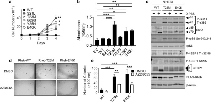
Fig. 3.
Rheb mutants drive increased cell proliferation and growth in soft agar. a MTT assay data for HEK293 cells transiently expressing the indicated Rheb mutants in medium lacking FBS. Cell number is calculated against a standard curve generated by 1:2 serial dilution of HEK293 cells. b BrdU incorporation assay for HEK293 cells transiently expressing the indicated Rheb mutants. Growth medium was replaced with medium lacking FBS for 24 h prior to analysis. BrdU was allowed to be incorporated for 2 h. c Western blot analysis of monoclonal NIH3T3 cells stably over-expressing Rheb-WT, T23M or E40K. Cell medium was replaced with medium either containing or lacking FBS 16 h and then replaced with D-PBS as indicated for 1 h prior to harvest. d Colony formation assay for NIH3T3 cells stably over expressing Rheb-WT, T23M or E40K. Liquid medium was replaced twice per week with medium containing 1 µM AZD8055 or DMSO as indicated. Number of colonies larger than 100 nM were counted by hand and are quantified in (e). For panels a, b and e, n = 3 (means ± S.D.). Statistical significance was determined by Student’s t-test where *0.01 ≤ P < 0.05; **0.001 ≤ P < 0.01; ***P < 0.001

