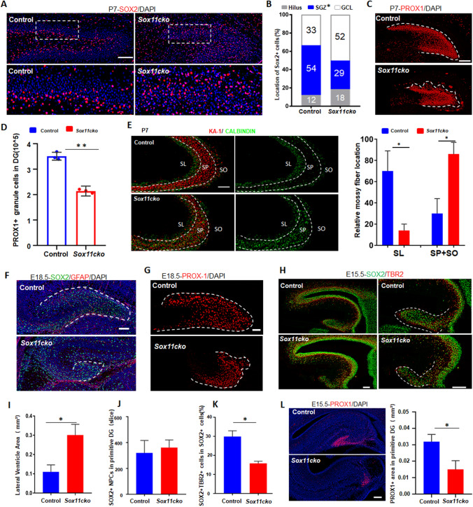
Fig. 3.
Pathological defects in the SOX11-deficient hippocampus are initiated from abnormal progenitor polarity and defective differentiation in the E13.5–E15.5 embryonic brain. A Staining of SOX2 + NPCs in the postnatal dentate gyrus at P7. Scale bar, 100 μm. B Distribution of SOX2 + NPCs in the postnatal dentate gyrus in the P7 brain from the SOX11-mutant or control mice. GCL, SGZ and Hilus. n = 3 mice, *p < 0.01 (Student’s t test). Staining (C) and quantification (D) of PROX1 + granule cells at the DG in P7 brain from SOX11-mutant or control mice. Scale bar, 100 μm. E Mossy fiber connections decreased to the SL region but increased to the SP and SO regions in the SOX11-deficient hippocampal CA3 region at P7. Scale bar, 100 μm, Error bars, mean ± SEM, n = 3, *p < 0.05, (Student’s t test). Immunostaining of SOX2 + NPCs (F) and PROX1 + granule cells (G) at the primitive dentate gyrus in the E18.5 brain from the SOX11-mutant or control mice. Scale bar, 50 μm. H and I SOX11 deficiency leads to decreased primitive DG region and enlarged ventricles in the developing primitive dentate gyrus in the E15.5 mouse brain. Scale bar, 200 μm. Error bars, mean ± SEM, n = 3 mice, *p < 0.05, (Student’s t test). The number of SOX2 + cells did not show reduction (J), but the percentage of double-positive cells (SOX2 and TBR2) in total SOX2 + cells (K) was significantly decreased in the developing primitive dentate gurus in the E15.5 mutant hippocampus. Error bars, mean ± SEM, n = 3 mice, *p < 0.05, (Student’s t test). L The area of PROX1 + granule cells decreased significantly in the developing primitive dentate gyrus in the E15.5 mutant hippocampus. Scale bar, 200 μm. Error bars, mean ± SEM, n = 3 mice. *p < 0.05, (Student’s t test)

