Fig. 13.
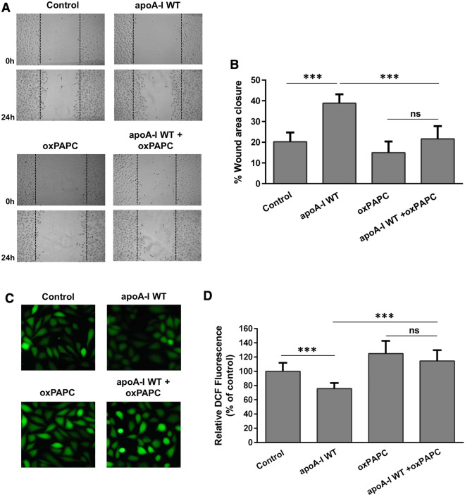
Migration and ROS formation by EA.hy926 cells incubated with lipoprotein particles containing WT apoA-I in the absence or presence of oxPAPC. a EA.hy926 cells were wounded and treated with or without rHDL containing WT apoA-I, in the absence or presence of oxPAPC, for 24 h. Representative microscopic views at 0 h and 24 h are shown. Images were scanned and quantified by ImageJ software. b Quantitative analysis of wound area closure for EA.hy926 cells after 24 h. Values are the means ± SD (n = 6). c EA.hy926 cells treated with or without rHDL containing WT apoA-I, in the absence or presence of oxPAPC, for 24 h, were then incubated with DCFH-DA. The formation of ROS by cells was measured following detection of fluorescent DCF emitted from cells using a fluorescence microscope. Representative microscopic views are shown. Images were scanned and quantified by Image J software. d The relative DCF fluorescence is shown. Values are the means ± SD (n = 10). ***p < 0.0001

