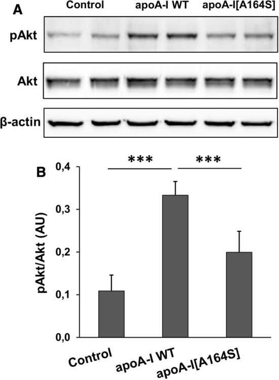
Fig. 9.

Activation of Akt kinase in EA.hy926 cells incubated with lipoprotein particles containing WT apoA-I or apoA-I[A164S]. Protein expression levels of pAkt, total Akt and β-actin in EA.hy926 cells left untreated or treated with rHDL containing WT apoA-I or apoA-I[A164S] for 24 h were measured by immunoblotting. a A representative set of immunoblots is shown. b Western blots were scanned and quantified by ImageJ software. The normalized levels of pAkt against total Akt are shown. Values are the means ± SD for four independent experiments performed in duplicate. AU arbitrary units. ***p < 0.0001
