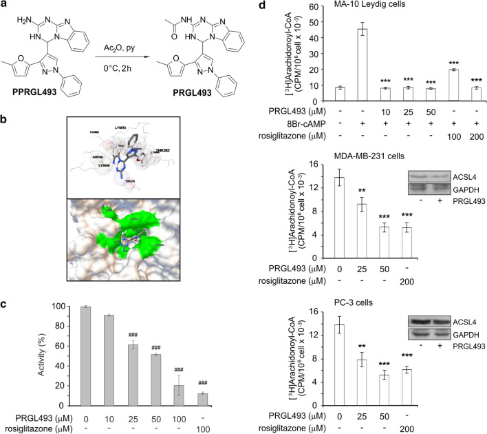
Fig. 2.
PRGL493 inhibits ACSL4 activity. a Chemical structure of PPRGL493 (4-(3-(5-methylfuran-2-yl)-1-phenyl-1H-pyrazol-4-yl)-3,4-dihydrobenzo[4,5]imidazo[1,2-a][1,3,5]triazin-2-amine) in a scheme showing the synthesis of PRGL493 (N-(4-(3-(5-methylfuran-2-yl)-1-phenyl-1H-pyrazol-4-yl)-3,4-dihydrobenzo[4,5]imidazo[1,2-a][1,3,5]triazin-2-yl)acetamide). b A modelling of 3D structure of human ACSL4 (isoform 2, NCBI Reference Sequence: NP_001305438.1) and compound PPRGL493 showing the best docking of compound PPRGL493 in balls-and-sticks representation and the residues in close contact with ACSL4 as spheres; a hydrogen bond is indicated by the green dotted line. A hACSL4 model surface with compound PPRGL493 in the AA tunnel entrance is shown below. c Inhibition of recombinant ACSL4. Recombinant flag-tagged hACSL4 was incubated in the presence or absence of inhibitors, i.e. increasing concentrations of PRGL493 (up to 100 μM) or rosiglitazone (100 μM) for 10 min at 37 °C. Acyl-CoA synthetase activity was measured as AA-CoA formation from AA following the protocol described in Materials and Methods. Data are presented as a percentage of total enzymatic activity. Data represent the means ± SD. ###p < 0.001 vs. total enzymatic activity without inhibitors. d Inhibition of ACSL4 activity in MDA-MB-231, PC-3 and MA-10 Leydig cells. Cell lines were incubated with [3H]-AA (0.5 µCi/ml in serum-free medium) for 3 h were treated with inhibitor PRGL493 at different concentrations (0, 25 and 50 μM) or rosiglitazone (200 μM) for 3 h and serum was later added for 48 h. MA-10 Leydig cells were treated with inhibitor PRGL493 at different concentrations (0, 10, 25 and 50 μM) or rosiglitazone (100 and 200 μM) for 3 h and then stimulated with 8Br-AMPc (0.5 mM) for 1 h. After treatments, [3H]AA-CoA formation was assessed and expressed as CPM/106 cells × 10–3. Data represent the means ± SD. **p < 0.01 and ***p < 0.001 vs. control cells (in the absence of inhibitors). Western blot analysis of ACSL4 expression in MDA-MB-231 and PC-3 cells treated with PRGL493 (50 μM) is shown as respective insets

