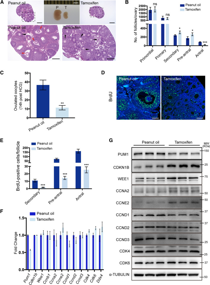
Fig. 5.
Tamoxifen-induced Pum1 knockout in the background of Pum2−/− mice resulted in follicle maturation defects. A Hematoxylin and eosin-stained ovarian sections showed that ovaries of tamoxifen-treated mice contained few antral follicles. Peanut oil-treated littermates of the same genotype (Ert2-cre/ + ; Pum1F/F; Pum2−/−) were used as controls. White arrow, secondary follicle; Black arrow, pre-antral follicle; Red arrow, antral follicle. Scale bar: 500 μm (upper panel); 200 μm (lower panel). B Quantitative analysis of primordial follicles, primary follicles, secondary follicles, pre-antral follicles, and antral follicles at three weeks of age. Mice were injected intraperitoneally with peanut oil or tamoxifen for five consecutive days (n = 3 for each group, data are mean ± SEM). ***P < 0.001; **P < 0.01; *P < 0.05; ns, not statistically significant. C Number of ovulated oocytes from Ert2-cre/ + ; Pum1F/F; Pum2−/− female mice treated with peanut oil (n = 3) or tamoxifen (n = 3). Oocytes were collected at 14 h after HCG. D Representative images of ovarian granulosa cell proliferation assay using BrdU labeling after peanut oil or tamoxifen induction. The incorporated BrdU was stained with an anti-BrdU monoclonal antibody (green), DAPI was applied as a nuclear counterstain (blue). Scale bar, 100 μm. E Quantification of BrdU-positive cells in secondary follicles, pre-antral follicles, and antral follicles, respectively. Each group consisted of three mice, and three discontinuous ovarian sections were analyzed for each Ert2-cre mouse. ***P < 0.001. F RT-qPCR analysis of mRNA expression of various cell cycle factors after intraperitoneal injection of peanut oil or tamoxifen. G Immunoblot analysis of PCCR protein expression in Ert2-cre/ + ; Pum1F/F; Pum2−/− female ovaries after treatment with tamoxifen or peanut oil

