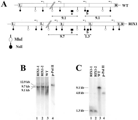
Figure 7.
Comparison of the structures of integrated new rDNA repeats and that of native rDNA repeats. (A) The structure of integrated new rDNA repeats (RIX1) and that of native rDNA repeats (WT) are schematically shown. The sizes of the L and R segments are not to scale. Expected sizes of fragments detected after digestion of genomic DNA with MluI or NsiI and hybridized with radioactive probe a or b (indicated as a filled box) are indicated (in kb). (B and C) Southern analysis of genomic DNA using MluI and probe a (B) and that using NsiI and probe b (C). DNA was isolated and analyzed from the following strains: two independent clones (RIX1-1 and RIX1-2) with integrated new rDNA repeats obtained after hygromycin selection, lanes 1 and 2; NOY505 (WT), lane 3; plasmid pNOY353 (p-Pol II), lane 4. The sizes of bands estimated from size markers run in parallel are indicated.

