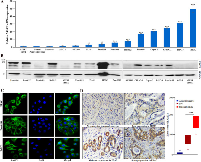
Fig. 2.
Overexpression of LAMC2 transcript and protein in PDAC cell lines and clinical samples. A Quantitative real-time PCR (qPCR) displayed upregulation of LAMC2 transcript in PDAC cells compared to normal human pancreatic tissue and cell line (hTERT-HPNE). Results represent mean ± SD; n = 3. **P-value < 0.001; ***P-value < 0.0001; two-tailed t-test. B Western blot analysis displayed higher LAMC2 protein expression in PDAC cell lines compared to hTERT-HPNE (normal human pancreatic epithelial) cells. C Immunofluorescence experiments demonstrated LAMC2 protein expression in the cytoplasm of PDAC cells. DAPI (4′,6′-diamino-2-phenylindole) was used for nuclear staining. D IHC (immunohistochemistry) data showed strong, moderate, and low staining for LAMC2 in the representative PDAC clinical tissue sections. No/negative staining was noticed in normal pancreatic tissue sections (original magnification, X200). Histoscore analysis displayed significant difference in the staining intensity among low and moderate/high groups. Results represent mean ± SD. ***P-value < 0.0001

