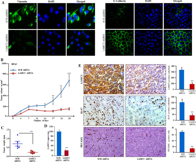
Fig. 5.
Inhibition of LAMC2 suppressed the tumor growth in PDAC xenografts. A Immunofluorescence staining of LAMC2 silenced cells displayed expression of E-cadherin on the cell surface and suppressed the expression of vimentin in PDAC cells. DAPI was used for nuclear staining. B Graph displayed decreased tumor volumes and growth in the LAMC2 silenced group compared to the scramble shRNA group (n = 6). C The weight of the tumors in the LAMC2 silenced group was significantly lower (**P-value < 0.001; t-test) than the scramble shRNA group. D qPCR confirmed LAMC2 knockdown in tumor samples. E IHC on tumor section displayed decreased reactivity for LAMC2, Ki-67, and PECAM1 in LAMC2 silenced tumors sections compared to scramble shRNA Tumor sections. Data represent mean ± SD; n = 3. **P-value < 0.001; two-tailed t-test. Original magnification, X200

