Fig. 5.
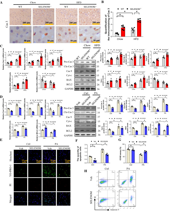
SELENOM−/− increases HFD-mediated mitochondrial pathway apoptosis in the liver. A, B Cas 3 was measured via immunohistochemistry analysis in livers from WT, SELENOM−/−, HFD and SELENOM−/− + HFD. Six fields (Scale bar: 200 and 50 μm) were randomly selected for each sample. The positive area in each image was measured (n = 6; *P < 0.05). C The mRNA and protein expression levels of mitochondrial pathway apoptosis-related genes Cas 3, Cas 9, Bax and Bcl2 were determined in HFD-treated livers with SELENOM−/− (n = 6; *P < 0.05). D The mRNA and protein expression levels of mitochondrial pathway apoptosis-related genes Cas 3, Cas 9, Bax and Bcl2 were determined in hepatocytes of Veh, SELENOM, PA and SELENOM + PA (n = 3; *P < 0.05). E, F Apoptosis and necrosis staining with YO-PRO-1 and PI for apoptosis detection, and the green nucleus represent an apoptotic cell (n = 3; *P < 0.05). Fields from one representative experiment of three are shown. G CCK8 assay was used to assess the hepatocyte viability under PA treatment (n = 3; *P < 0.05). H Cell viabilities and apoptosis rates were determined by flow cytometry. Healthy (Q1), early apoptotic (Q2), late apoptotic (Q3), and necrotic populations (Q4). Images were chosen from three independent biological samples (n = 3; *P < 0.05). Values represent means ± SEM

