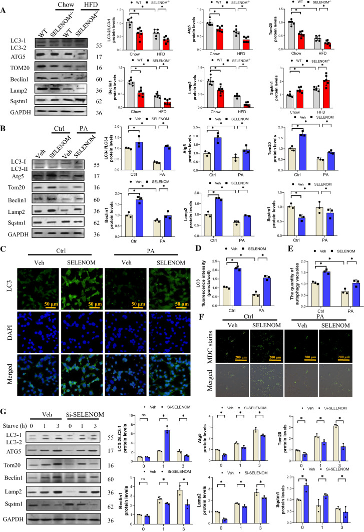
Fig. 6.
SELENOM involves in HFD-inhibited mitophagy in the liver. A The expression levels of mitophagy-related proteins such as LC3-I, LC3-II, Atg5, Tom20, Beclin1, Lamp2 and Sqstm1 in livers from WT, SELENOM−/−, HFD and SELENOM−/− + HFD (n = 6; *P < 0.05). B The expression levels of mitophagy-related proteins such as LC3-I, LC3-II, Atg5, Tom20, Beclin1, Lamp2 and Sqstm1 in hepatocytes of Veh, SELENOM, PA and SELENOM + PA (n = 3; *P < 0.05). C, D Immunofluorescence assay for mitophagy via assessing LC3 in vitro. Fields from one representative experiment of three are shown (n = 3; *P < 0.05). E, F The quantity of autophagy vacuoles was recorded by MDC staining. Fields from one representative experiment of three are shown (n = 3; *P < 0.05). G Immunoblotting for LC3-I, LC3-II, Atg5, Tom20, Beclin1, Lamp2 and Sqstm1 in AML12 hepatocytes treated with Si-NC or Si-SELENOM were subjected to amino acid starvation for the indicated durations, 0, 1, 3 h, respectively (n = 3; *P < 0.05). Values represent means ± SEM

