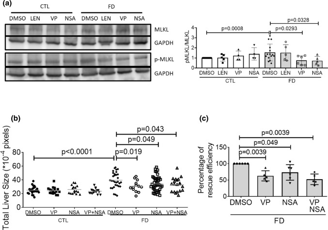
Fig. 9.
The lack of additive rescuing effects upon co-exposure to verteporfin and NSA for FD-induced hepatomegaly. a The cell lysates prepared from Huh7 cells cultivated in FD medium in the presence of LEN (TNFα inhibitor, 4 µM), VP (Hippo pathway inhibitor, 0.1 µM) or NSA (necroptosis inhibitor, 2 µM) for 8 days were analyzed with Western blotting for MLKL phosphorylation and quantified. b Larvae were exposed to VP (0.62 µM) and NSA (5 µM) either separately or simultaneously from 7 to 11 dpf. Larvae were imaged at 11 dpf and larval liver size was quantified with on-line software Image J. c VP (0.1 µM) and NSA (2 µM) were added to FD cultured medium for Huh7 cells separately or simultaneously 5-day post-seeding. Cell sizes were measured on 8 dps with flow cytometry and calculated for the percentage of rescuing efficiency. Shown here are the averaged results of at least three independent trials. LEN lenalidomide, VP verteporfin, NSA necrosulfonamide. Statistical results are represented in the mean ± SEM. CTL control (Tg-GGH/LR larvae without FD or Huh7 cells cultured in regular α-MEM), FD folate deficiency

